

Written by Julie Bick, Ph.D.
Mitochondria are most often referred to as the "powerhouses of the cell," and play a crucial role in generating the energy required for various cellular functions. However, their influence extends far beyond mere energy production, and research is linking mitochondrial health with our biological aging as well as our susceptibility to many common diseases. In this blog we review the multifaceted roles of mitochondria in cellular health and aging, exploring their functions and the mechanisms by which they contribute to the aging process, and their emerging roles in disease onset and progression.
Mitochondria are double-membrane-bound organelles found in the cytoplasm of nearly all eukaryotic cells. Their unique structure is key to their functionality. The outer membrane is permeable to ions and small molecules, allowing the exchange of substances between the cytoplasm and the intermembrane space, in which various biochemical processes occur. The inner membrane is impermeable to most molecules and houses the protein complexes of the electron transport chain (ETC). It is highly folded into structures called cristae, which increase its surface area, enhancing the organelle's ability to produce ATP. The innermost compartment of the mitochondrion contains enzymes, mitochondrial DNA (mtDNA), and ribosomes. The matrix is the site of the tricarboxylic acid (TCA) cycle (also known as the Krebs cycle or citric acid cycle) and other metabolic processes. All of these features are summarized below in Fig. 1.
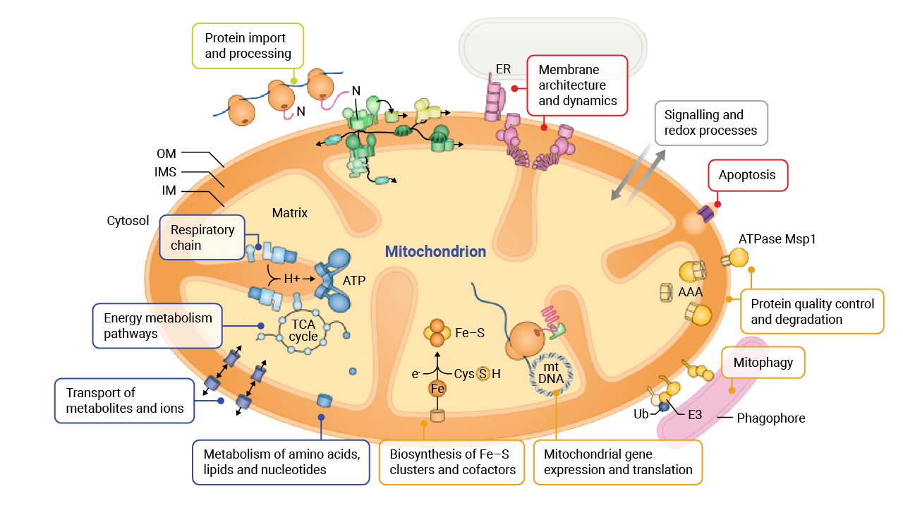
Fig. 1. Summary of the structure and multifaceted functions of mitochondria and their role in cellular metabolic pathways.
Mitochondria are best known for their role in energy production through oxidative phosphorylation, that involves electron-transport-linked phosphorylation or terminal oxidation reactions to generate ATP, the primary energy currency of the cell. This process involves the ETC and the enzyme ATP synthase, which together convert energy from nutrients into ATP. Aside from energy production, mitochondria are involved in various other metabolic pathways, including the TCA cycle, fatty acid oxidation, and amino acid metabolism. These pathways produce intermediates crucial for cellular functions and biosynthesis. In the case of fatty acid synthesis, mitochondria generate a molecule known as lipoic acid, a key cellular antioxidant molecule, a building block for lipids as well as a cofactor for several important mitochondrial enzyme complexes involved in ATP production and amino acid metabolism. There is increasing interest in lipoic acid’s potential to manage neurodegenerative diseases such as Alzheimer's as well as multiple sclerosis and type 2 diabetes- we’ll cover the potential therapeutic benefits of lipoic acid supplements in a later blog.
Another key role that mitochondria play is in the regulation of intracellular calcium levels, which are vital for numerous cellular processes, from muscle contraction, to neurotransmitter release, and cell signaling. In addition, mitochondria release pro-apoptotic factors, such as cytochrome c, in response to cellular stress, initiating the cascade that leads to apoptosis. For many years, researchers understood that disruptions in mitochondrial function could lead to various diseases as well as impact biological aging, and research is now describing the mechanisms for these changes.
The role of mitochondria in aging has been a subject of intense research and debate, and in recent years several theories have been proposed to explain how mitochondrial dysfunction contributes to biological aging.
The mitochondrial free radical theory of aging, first proposed by Dr. Denham Harman in the 1950s, suggests that accumulated damage from ROS produced during mitochondrial oxidative phosphorylation is a primary driver of aging (review Sanz A, Stefanatos RK. 2008). According to this theory the Reactive Oxygen Species (ROS) production by mitochondria as byproducts of ATP synthesis can damage mitochondrial DNA (mtDNA), proteins, and lipids. While ROS play key roles in cell signaling and immune system defense mechanisms, excessive ROS can damage cellular components. Unlike nuclear DNA, mtDNA is located close to the site of ROS production and lacks protective histones, making it more susceptible to oxidative damage. Healthy mitochondria are equipped with antioxidant systems to detoxify ROS, but if these defenses are not sufficient, then mitochondrial function can start to diminish. As the mtDNA is exposed to more ROS, mutations and deletions in mtDNA begin to impair mitochondrial function, leading to decreased ATP production and increased ROS generation, creating a vicious cycle of damage. This accumulation of mitochondrial damage leads to cellular dysfunction, reduced energy production, and increased oxidative stress, all of which further contribute to the aging process and age-related diseases.
Mitochondria are dynamic organelles that undergo continuous cycles of fusion and fission, processes collectively known as mitochondrial dynamics. Together these processes are crucial for maintaining mitochondrial function and cellular health.
Mitochondrial fusion involves the merging of two mitochondria, allowing for the mixing of mitochondrial contents, including mtDNA and proteins. The theory is that this process helps dilute damaged components and maintain mitochondrial function.
In contrast, mitochondrial fission involves the division of a single mitochondrion into two separate organelles. Fission is important for removing damaged mitochondria through a process called mitophagy, a specialized form of autophagy that targets dysfunctional mitochondria for degradation. It involves the selective autophagic degradation of mitochondria, preventing the accumulation of damaged organelles that could compromise cellular function. During this process, key mitochondrial components are recycled to form new healthy mitochondria. In this way, mitophagy plays a central role in adjusting mitochondrial numbers (known as the mitochondrial load or mass) to changing metabolic needs of cells during growth or tissue healing for example (Picca et. al. 2023). Given this critical role, it isn’t surprising that dysregulated mitophagy has been implicated in various age-related diseases, including neurodegenerative disorders and cardiovascular diseases.
In addition, other quality control mechanisms for mitochondria have more recently been described; these include Mitochondrial-Lysosome-Related Organelles (MLRO) that to date have been most extensively studied in the liver. They appear to represent the primary pathway for mitochondrial degradation in the dedifferentiated hepatocytes commonly associated with the late stage of chronic liver diseases such as ALD and MASH, which leads to liver failure (Ma et. al. 2023). Cellular stress has been shown to induce the formation of Mitochondrial Derived Vesicles (MDV) that are enriched in oxidized mitochondrial proteins (Neuspiel et. al. 2008). Following fusion with lysosomes, these vesicles and their contents are degraded. Significantly, MDVs have been demonstrated to protect against hypoxia-induced cardiomyocyte damage, as well as play a potential role in the inflammation responses associated with the progression of Parkinson’s Disease (Matheoud et. al. 2016).
Mitochondrial dysfunction has long been seen as a key player in aging, and now by deciphering the specific roles mitochondria play in the development and progression of age-related conditions, we can propose mitochondrial-focused therapeutic strategies.
Neurodegenerative diseases, such as Alzheimer's disease, Parkinson's disease, and amyotrophic lateral sclerosis (ALS), are all characterized by the progressive loss of neurons that correlates with mitochondrial dysfunction. In the example of Alzheimer's Disease, mitochondrial dysfunction associated with impaired energy metabolism, increased oxidative stress, and the accumulation of amyloid-beta plaques.
In Parkinson's Disease, mitochondrial dysfunction is linked to the loss of dopaminergic neurons in the substantia nigra. Mutations in genes associated with mitochondrial function, such as PINK1 and Parkin, have been implicated in disease onset and progression, and therefore enhancing mitophagy and improving mitochondrial quality control are potential therapeutic targets. A recent study identified the presence of cell-free mtDNA with deletions in cerebrospinal fluid. It is proposed that this modified mtDNA could serve as an early marker for severe brain diseases such as Parkinson’s Disease. This discovery is crucial because it suggests a potential initial link in the neurodegenerative process, which eventually leads to the motor and cognitive symptoms seen in disorders like Parkinson’s disease and Lewy body dementia (Puigròs et. al. 2024).
Mitochondrial dysfunction is also prominent in ALS, a devastating neurodegenerative disease characterized by the progressive loss of motor neurons. Sadly, there are currently very limited treatment options. It is now well characterized that ALS is associated with impaired energy production, increased oxidative stress, and mtDNA damage (Zhao et. al. 2022), and so therapeutic strategies aimed at protecting mitochondrial function and reducing oxidative stress are being investigated. These include the use of dichloroacetate, ketogenic and high-fat diets, acetyl-carnitine, and mitochondria-targeted antioxidants (Cunha-Oliveira, et. al. 2024).
Cardiovascular diseases, including heart failure, ischemic heart disease, and hypertension, are major contributors to morbidity and mortality in aging populations. Since cardiac tissue sources most of its energy through oxidative metabolism, disruptions in these mitochondrial pathways are thought to be a primary mechanism linking mitochondrial dysfunction and failure in contractile performance of the heart. However, the role of mitochondria in heart failure is now increasingly recognized to extend beyond this and new evidence is highlighting a vicious cycles of pathophysiological mechanisms involving metabolic flux, protein modification/oxidation, ROS-induced ROS generation leading to redox imbalance, impaired mitochondrial Ca2+ homeostasis, and inflammation. The hope is that understanding how these all develop in heart disease will lead to novel avenues for therapeutic interventions (Zhou and Tian, 2018).
Metabolic disorders, including obesity, diabetes, and fatty liver disease, are closely linked to mitochondrial dysfunction. Both functional and structural changes in mitochondria have been described in association with these conditions. As such, there has been increasing focus on the biogenesis of mitochondria as well as increased expression of antioxidant enzyme systems as potential therapies. Several pharmacological strategies to trigger these responses have been proposed such as bezafibrate to activate the PPAR-PGC-1α axis, resveratrol and quercetin to activate AMPK and act as Sirt1 agonists respectively, as well as the adoption of antioxidant supplements such as coenzyme Q10, MitoQ10 and other mitochondria-targeted antioxidants including N-acetylcysteine (NAC), vitamin C, vitamin E vitamin K1, vitamin B, sodium pyruvate or -lipoic acid. Only long-term studies will help highlight the success of these interventions in managing disease progression (Valero, 2014).
Mitochondria are essential organelles that play a central role in cellular health and aging. Their functions extend far beyond energy production, encompassing metabolic regulation, calcium signaling, apoptosis, and ROS detoxification. Mitochondrial dysfunction is a hallmark of aging and is implicated in the development of various age-related diseases, including neurodegenerative disorders, cardiovascular diseases, and metabolic disorders. Understanding the mechanisms by which mitochondria influence cellular health and aging can provide insights into potential therapeutic strategies. Interventions aimed at improving mitochondrial function, such as caloric restriction, exercise, and pharmacological treatments, hold promise for promoting healthy aging and reducing the burden of age-related diseases.
As research continues to reveal the complexities of mitochondrial biology, the potential for targeted therapies to enhance mitochondrial function and improve health-span and lifespan becomes increasingly promising. Long-gone are the days when we only refer to mitochondria as the energy powerpacks of the cell. It is now accepted that these organelles represent both the guardians of cellular health and drivers of aging and remain a fascinating and crucial area of study in biological aging and the progression of a range of diseases.
In our next blog we’ll review the evidence for novel nutraceutical approaches to maintain mitochondrial health, and explore some of the techniques that researchers are employing to assess their influence on our overall health and aging.